Leg Bones Diagram - Ballet Feet | ARHtistic License. The femur, or thigh bone, is the largest, heaviest, and strongest bone in the human body. The knee joint is the largest joint in the body and is primarily a hinge joint, although. Next to the tibia is the fibula the thinner weaker bone of the lower leg. The human leg, in the general word sense, is the entire lower, and footlimb of the human body, including the foot, thigh and even the hip or gluteal region. At the same time, the bones and joints of the leg and foot must be strong enough to support the body's weight while remaining flexible enough for movement and balance.
Distal end of right humerus. Other sets by this creator. Next to the tibia is the fibula the thinner weaker bone of the lower leg. While their parts are similar in general, their structure has been adapted to differing functions. The knee joint is the largest joint in the body and is primarily a hinge joint although some sliding and rotation occur.
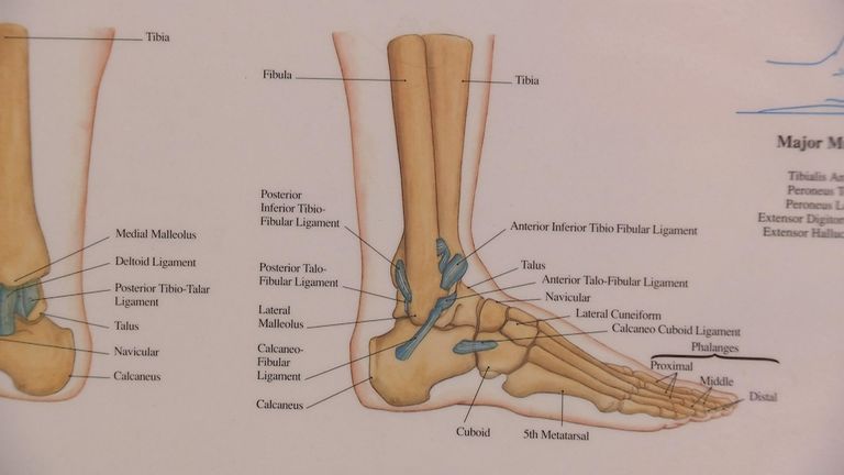
Labeled Diagram of the Human Leg by xKeren on DeviantArt from img00.deviantart.net We'll break down the anatomy and function of the upper leg, knee, lower leg, ankle, and foot. Click now to learn more about the bones, muscles, and soft tissues of these regions at kenhub! Human leg bones vector image. The foot bones shown in this diagram are the talus, navicular, cuneiform, cuboid, metatarsals and calcaneus. While their parts are similar in general, their structure has been adapted to differing functions. Human foot bones anatomy sketch of orthopedics medicine. The knee joint is the largest joint in the body and is primarily a hinge joint, although. It expands at the proximal and distal ends, articulating at the knee and ankle joints respectively.
The bones of your leg have roughened patches on their surfaces where muscles are attached.
When your muscles contract, they pull the bone they're. Diagram of blood and nerve supply to bone. Human foot bones anatomy sketch of orthopedics medicine. Skeleton leg ankle joints and toe phalanges, cuboid, metatarsal, navicular and cuneiform bones, hand drawn dorsal view of foot. Distal end of right humerus. Human leg bones vector image. Health diagram bone skeleton leg knee science anchor chart human human body. However, the definition in human anatomy refers only to the section of the lower limb extending from the knee to the ankle, also known as the crus. Want to learn more about it? It expands at the proximal and distal ends, articulating at the knee and ankle joints respectively. Time to jump right into the biggest and strongest bones in the human body. Your leg bones are the longest and strongest bones in your body. This diagram depicts diagram leg bones anatomy.
At the same time, the bones and joints of the leg and foot must be strong enough to support the body's weight while remaining flexible enough for movement and balance. High resolution textures and displacement included. Blood vessels and nerves enter the bone through the nutrient foramen. This diagram depicts diagram leg bones anatomy. The bones and features labelled are the femur, patella, fibula.
Luke Shaw expected to be out for up to nine months, expert says | Football News | Sky Sports from e2.365dm.com Your leg bones are the longest and strongest bones in your body. The musculoskeletal segment of the leg, including the foot bones (ankle, heel bone, toe bones), fibula and tibia, knee, femur and femoral neck, hip and sacrum as well as the third, fourth, and fifth lumbar vertebrae (l3, l4, l5) are a functional unit. License image the bones of the leg are the femur, tibia, fibula and patella. The foot bones shown in this diagram are the talus, navicular, cuneiform, cuboid, metatarsals and calcaneus. Joints of hand anterior view, lateral view, right hand. High quality realistic skeleton legs. The bones of your leg have roughened patches on their surfaces where muscles are attached. Human leg bones vector image.
The foot bones shown in this diagram are the talus, navicular, cuneiform, cuboid, metatarsals and calcaneus.
It expands at the proximal and distal ends, articulating at the knee and ankle joints respectively. File is ready to render. These can include any the following: Human anatomy diagrams show internal organs, cells, systems, conditions, symptoms and sickness information and/or tips for healthy living. Includes leg (femur, tibia, patella, and fibula) and foot (tarsals and digits) bones. Blood vessels and nerves enter the bone through the nutrient foramen. Diagram of blood and nerve supply to bone. Distal end of right humerus. When your muscles contract, they pull the bone they're. Your leg bones are the longest and strongest bones in your body. The bones of the leg are the femur, tibia, fibula and patella. Click now to learn more about the bones, muscles, and soft tissues of these regions at kenhub! High resolution textures and displacement included.
The musculoskeletal segment of the leg, including the foot bones (ankle, heel bone, toe bones), fibula and tibia, knee, femur and femoral neck, hip and sacrum as well as the third, fourth, and fifth lumbar vertebrae (l3, l4, l5) are a functional unit. Visit kenhub for more skeletal system quizzes. Cheek bone (zygoma) upper jaw (maxilla). Your legs are two of your most important body parts. The foot bones shown in this diagram are the talus, navicular, cuneiform, cuboid, metatarsals and calcaneus.
Anatomy & Physiology Illustration from www.anatomyfacts.com High quality realistic skeleton legs. Disposition of rotator cuff muscles diagram. Human anatomy diagrams show internal organs, cells, systems, conditions, symptoms and sickness information and/or tips for healthy living. A leg bone is a bone found in the leg. It mainly serves as an attachment point for the muscles of the lower leg. Want to learn more about it? The human leg, in the general word sense, is the entire lower, and footlimb of the human body, including the foot, thigh and even the hip or gluteal region. Quizzes on human skeletal system anatomy, bone anatomy, and bone markings.
Disposition of rotator cuff muscles diagram.
High resolution textures and displacement included. Includes obj for maximum compatibility. Cheek bone (zygoma) upper jaw (maxilla). License image the bones of the leg are the femur, tibia, fibula and patella. This diagram shows the bones of the femur and the patella. The musculoskeletal segment of the leg, including the foot bones (ankle, heel bone, toe bones), fibula and tibia, knee, femur and femoral neck, hip and sacrum as well as the third, fourth, and fifth lumbar vertebrae (l3, l4, l5) are a functional unit. Visit kenhub for more skeletal system quizzes. Includes leg (femur, tibia, patella, and fibula) and foot (tarsals and digits) bones. Quizzes on human skeletal system anatomy, bone anatomy, and bone markings. However, the definition in human anatomy refers only to the section of the lower limb extending from the knee to the ankle, also known as the crus. The foot bones shown in this diagram are the talus, navicular, cuneiform, cuboid, metatarsals and calcaneus. The bones and features labelled are the femur, patella, fibula. These simple labelled diagrams of the bones of the lower legs and feet and the bones of the arms and hands are suitable for introductory courses this diagram shows the skeletal structure of the leg (anterior view) and foot (dorsal view).
Share :
Post a Comment
for "Leg Bones Diagram - Ballet Feet | ARHtistic License"


Post a Comment for "Leg Bones Diagram - Ballet Feet | ARHtistic License"